
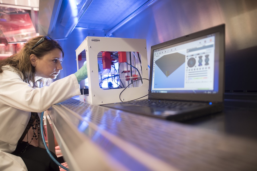
Dr. Angela Mortari uses a bioprinter to study spreading tumors. Photo courtesy of the University of Minnesota.
In today's cancer research world, ink can be made out of biological materials, and human tissue can be printed three-dimensionally.
Angela Mortari, PhD, vice chair for research, professor in the Department of Pediatrics at the University of Minnesota Medical School and director of the 3D Bioprinting Facility, is at the forefront of this innovative technology, using a 3D bioprinter to explore and follow the behaviors of fatal cancer cells.
"Think of the average person walking in the city and how they behave and interact," said Dr. Mortari. "Now imagine placing that person up in the tundra, completely naked. You'll see glimpses of their normal behavior, like walking, but you'll also see very exaggerated behaviors, like shivering, and none of their daily routine."
This is how a cancer cell reacts in a two-dimensional petri dish when doused with cancer treatments: exaggerated, and oftentimes, largely unlike how the cell would normally behave in the human body.
"Two-dimensional models are just not good representations [of cancer]," said Dr. Mortari. "When you have a 3D soft-tissue model, you have a more normal context, and you make your research results more meaningful."
The idea that a tumor's surrounding biological environment could affect its behavior has existed for decades, but few institutions have tried to build 3D technology to study the concept.
That's why, about five years ago, Dr. Mortari sought to bring state-of-the-art 3D bioprinting technology to the University of Minnesota. She believed that cancer cells would behave more like they did in the human body with an environment made out of the same biomaterials found in the body, thus creating more relevant cancer research.
Knowing that the right talent existed at the University of Minnesota, Dr. Mortari cobbled together enough funding to bring biomedical and mechanical engineers, surgeons and stem cell scientists (among many others) on board and purchased the 3D-bioprinting technology and generated the biomaterials.
With her colleagues Michael C. McAlpine, PhD, and Faben Meng, PhD, from the College of Science and Engineering at the University of Minnesota, Dr. Mortari custom-built a 3D printer to create a tumor with a blood vessel supply (i.e., vascularized tumor tissues) in the context of other cell types typically found in a tumor. Her team precisely placed living cells, biomaterials and programmable release capsules within the tissues to imitate what they believe happens in the body as cancer spreads.
Using the 3D bioprinter, Dr. Mortari and her colleagues created an environment that caused the cancer cells to travel from a tumor through a blood vessel, becoming a metastatic (or travelling) cancer cell. In real time, the research team could see just how the cells spread in the vessels, and they confirmed that cancer cells behave much differently in a soft-tissue environment than in other kinds of models with glass or plastic surfaces.
In their bioprinted model, the cancer cells also grew more slowly than in a petri dish, mirroring what researchers think happens in the human body. Because of how the model is constructed, they were able to collect the metastatic cancer cells in a separate chamber and analyze them without destroying the printed model. This allows for the study of these circulating tumor cells to understand why they decided to enter the blood vessel and metastasize.
With this new knowledge, Dr. Mortari and her colleagues can identify effective potential therapies and test anticancer treatments for metastatic cancer in a more targeted, controlled way because the model allows for the introduction of drugs through the blood vessel that was created.
So far, Dr. Mortari and her colleagues have focused on studying lung cancer and melanoma, but she hopes to tackle a variety of cancers in the near future. Next, she plans to incorporate more cell types and cell therapies in the tumor models and cell therapies in the tumor models and study how they interact.
Support Leading Cancer Researchers
Children’s Cancer Research Fund invests in the brightest minds in cancer research, like Dr. Mortari. Your donation is the fuel researchers need to get them to the next big breakthrough in childhood cancer treatments.